Evaluation of the osteogenic potential and vascularization of 3D poly(3)hydroxybutyrate scaffolds subcutaneously implanted in nude rats.
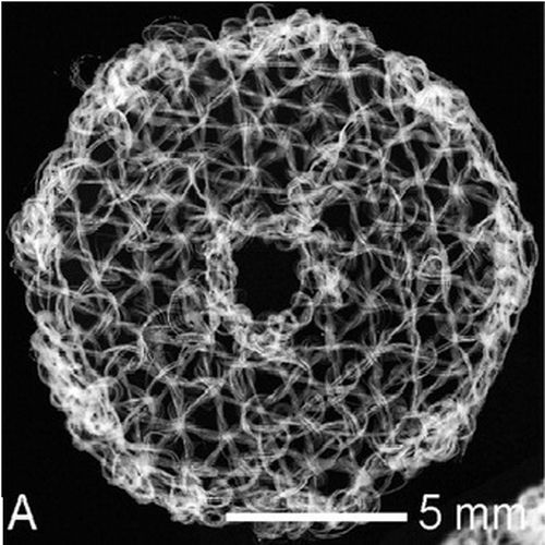
The aim of this study was to evaluate the osteogenic potential and the vascularization of embroidered, tissue engineered, and cell-seeded 3D poly(3)hydroxybutyrate (PHB) scaffolds in nude rats. Collagen I (coll I)- and collagen I/chondroitin sulfate (coll I/CS)-coated PHB scaffolds were seeded with human mesenchymal stem cells (hMSCs). Proliferation and differentiation were characterized by different biochemical assays in vitro. For animal experiments, the cells were cultivated on coll I- or coll I/CS-coated scaffolds and either expanded or osteogenically differentiated. Scaffolds were piled up to create a 3D scaffold pad and implanted subcutaneously into nude rats. In vitro hMSC showed proliferation and differentiation on PHB scaffolds. Alkaline phosphatase (ALP) and calcium increased in the differentiation medium and in the presence of coll I/CS. In vivo blood vessels were found in the scaffold-stack. Histological/immunohistological analyses of explanted scaffolds showed osteogenic markers such as osteopontin, osteonectin, and coll I around the PHB fibers. Coll I/CS-coated scaffolds with expanded hMSC showed higher values of ALP and calcium than the other combinations. Embroidered PHB scaffolds, coated with extracellular matrix components, provided an adequate environment and, therefore, a template for hMSC which could be differentiated in osteogenic direction.

- J Biomed Mater Res A 2010 Jan;92(1):185-95
- 2010
- Medical Biology
- 19170159
- PubMed
Enabled by:
