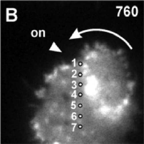
Propagating waves separate two states of actin organization in living cells.
Propagating actin waves are dynamic supramolecular structures formed by the self-assembly of proteins within living cells. They are built from actin filaments together with single-headed myosin, the Arp23 complex, and coronin in a defined three-dimensional order. The function of these waves in structuring the cell cortex is studied on the substrate-attached surface of Dictyostelium cells by the use of total internal reflection fluorescence (TIRF) microscopy. Actin waves separate two areas of the cell cortex from each other, which are distinguished by the arrangement of actin filaments. The Arp23 complex dominates in the area enclosed by a wave, where it has the capacity of building dendritic structures, while the proteins prevailing in the external area, cortexillin I and myosin-II, bundle actin filaments and arrange them in antiparallel direction. Wave propagation is accompanied by transitions in the state of actin with a preferential period of 5 min. Wave generation is preceded by local fluctuations in actin assembly, some of the nuclei of polymerized actin emanating from clathrin-coated structures, others emerging independently. The dynamics of phase transitions has been analyzed to provide a basis for modeling the nonlinear interactions that produce spatio-temporal patterns in the actin system of living cells.

- HFSP J 2009 Dec 30;3(6):412-27
- 2009
- Biophysics
- 20514132
- PubMed
Enabled by:
