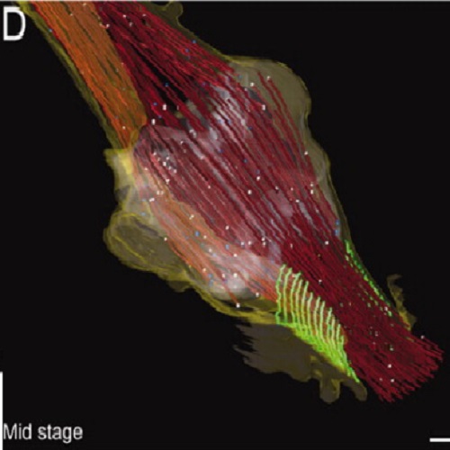
Cortical constriction during abscission involves helices of ESCRT-III-dependent filaments.
After partitioning of cytoplasmic contents by cleavage furrow ingression, animal cells remain connected by an intercellular bridge, which subsequently splits by abscission. Here, we examined intermediate stages of abscission in human cells by using live imaging, three-dimensional structured illumination microscopy, and electron tomography. We identified helices of 17-nanometer-diameter filaments, which narrowed the cortex of the intercellular bridge to a single stalk. The endosomal sorting complex required for transport (ESCRT)-III co-localized with constriction zones and was required for assembly of 17-nanometer-diameter filaments. Simultaneous spastin-mediated removal of underlying microtubules enabled full constriction at the abscission site. The identification of contractile filament helices at the intercellular bridge has broad implications for the understanding of cell division and of ESCRT-III-mediated fission of large membrane structures.

- Science 2011 Mar 25;331(6024):1616-20
- 2011
- Cell Biology
- 21310966
- PubMed
Enabled by:
