Elucidating membrane structure and protein behavior using giant plasma membrane vesicles.
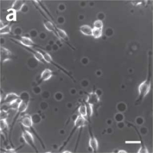
The observation of phase separation in intact plasma membranes isolated from live cells is a breakthrough for research into eukaryotic membrane lateral heterogeneity, specifically in the context of membrane rafts. These observations are made in giant plasma membrane vesicles (GPMVs), which can be isolated by chemical vesiculants from a variety of cell types and microscopically observed using basic reagents and equipment available in any cell biology laboratory. Microscopic phase separation is detectable by fluorescent labeling, followed by cooling of the membranes below their miscibility phase transition temperature. This protocol describes the methods to prepare and isolate the vesicles, equipment to observe them under temperature-controlled conditions and three examples of fluorescence analysis: (i) fluorescence spectroscopy with an environment-sensitive dye (laurdan); (ii) two-photon microscopy of the same dye; and (iii) quantitative confocal microscopy to determine component partitioning between raft and nonraft phases. GPMV preparation and isolation, including fluorescent labeling and observation, can be accomplished within 4 h.

- Nat Protoc 2012 Jun 03;7(6):1042-51
- 2012
- Biophysics
- 22555243
- PubMed
Enabled by:
