Immunohistochemical localization and characterization of putative mesenchymal stem cell markers in the retinal capillary network of rodents.
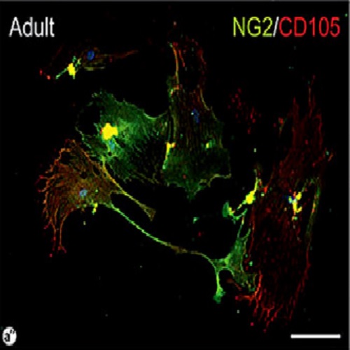
Perivascular cells of microvascular niches are the prime candidates for being a reservoire of mesenchymal stem cell (MSC)-like cells in many tissues and organs that could serve as a potential source of cells and a target of novel cell-based therapeutic approaches. In the present study, by utilising typical markers of pericytes (neuronal-glial antigen 2, NG2, a chondroitin sulphate proteoglycan) and those of MSCs (CD146 and CD105) and primitive pluripotent cells (sex-determining region Y-box 2, Sox2), the phenotypic traits and the distribution of murine and rat retinal perivascular cells were investigated in situ. Our findings indicate that retinal microvessels of juvenile rodents are highly covered by NG2-positive branching processes of pericytic (perivascular) cells that are less prominent in mature capillary networks of the adult retina. In the adult rodent retinal vascular bed, NG2 labeling is mainly confined to membranes of the cell body resulting in a pearl-chain-like distribution along the vessels. Retinal pericytes, which were identified by their morphology and NG2 expression, simultaneously express CD146. Furthermore, CD146-positive cells located at small arteriole-to-capillary branching points appear more intensely stained than elsewhere. Evidence for a differential expression of the two markers around capillaries that would hint at a clonal heterogeneity among pericytic cells, however, is lacking. In contrast, the expression of CD105 is exclusively restricted to vascular endothelial cells and Sox2 is detected neither in perivascular nor in endothelial cells. In dissociated retinal cultures, however, simultaneous expression of NG2 and CD105 was observed. Collectively, our data indicate that vascular wall resident retinal pericytes share some phenotypic features (i.e. CD146 expression) with archetypal MSCs, which is even more striking in dissociated retinal cultures (i.e. CD105 expression). These findings might have implications for the treatment of retinal pathologies.

- Cells Tissues Organs (Print) 2013 Apr 03;197(5):344-59
- 2013
- Medical Biology
- 23571553
- PubMed
Enabled by:
