Characterization of light lesion paradigms and optical coherence tomography as tools to study adult retina regeneration in zebrafish.
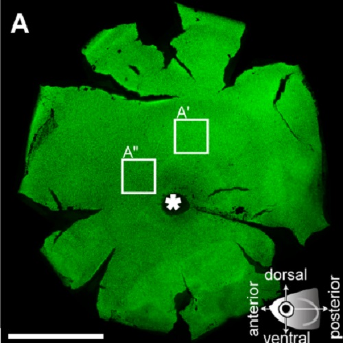
Light-induced lesions are a powerful tool to study the amazing ability of photoreceptors to regenerate in the adult zebrafish retina. However, the specificity of the lesion towards photoreceptors or regional differences within the retina are still incompletely understood. We therefore characterized the process of degeneration and regeneration in an established paradigm, using intense white light from a fluorescence lamp on swimming fish (diffuse light lesion). We also designed a new light lesion paradigm where light is focused through a microscope onto the retina of an immobilized fish (focused light lesion). Focused light lesion has the advantage of creating a locally restricted area of damage, with the additional benefit of an untreated control eye in the same animal. In both paradigms, cell death is observed as an immediate early response, and proliferation is initiated around 2 days post lesion (dpl), peaking at 3 dpl. We furthermore find that two photoreceptor subtypes (UV and blue sensitive cones) are more susceptible towards intense white light than red/green double cones and rods. We also observed specific differences within light lesioned areas with respect to the process of photoreceptor degeneration: UV cone debris is removed later than any other type of photoreceptor in light lesions. Unspecific damage to retinal neurons occurs at the center of a focused light lesion territory, but not in the diffuse light lesion areas. We simulated the fish eye optical properties using software simulation, and show that the optical properties may explain the light lesion patterns that we observe. Furthermore, as a new tool to study retinal degeneration and regeneration in individual fish in vivo, we use spectral domain optical coherence tomography. Collectively, the light lesion and imaging assays described here represent powerful tools for studying degeneration and regeneration processes in the adult zebrafish retina.

- PLoS ONE 2013 Nov 26;8(11):e80483
- 2013
- Neurobiology
- 24303018
- PubMed
Enabled by:
