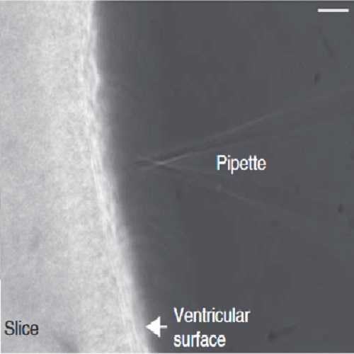
Microinjection of membrane-impermeable molecules into single neural stem cells in brain tissue.
This microinjection protocol allows the manipulation and tracking of neural stem and progenitor cells in tissue at single-cell resolution. We demonstrate how to apply microinjection to organotypic brain slices obtained from mice and ferrets; however, our technique is not limited to mouse and ferret embryos, but provides a means of introducing a wide variety of membrane-impermeable molecules (e.g., nucleic acids, proteins, hydrophilic compounds) into neural stem and progenitor cells of any developing mammalian brain. Microinjection experiments are conducted by using a phase-contrast microscope equipped with epifluorescence, a transjector and a micromanipulator. The procedure normally takes ?2 h for an experienced researcher, and the entire protocol, including tissue processing, can be performed within 1 week. Thus, microinjection is a unique and versatile method for changing and tracking the fate of a cell in organotypic slice culture.

- Nat Protoc 2014 May 24;9(5):1170-82
- 2014
- Neurobiology
- 24762784
- PubMed
Enabled by:
