Millisecond time resolution correlative light and electron microscopy for dynamic cellular processes.
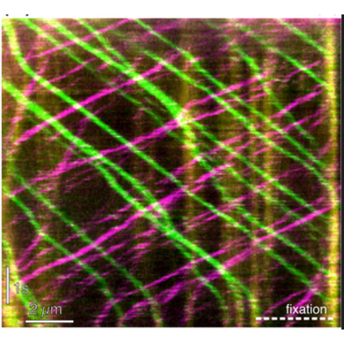
Molecular motors propel cellular components at velocities up to microns per second with nanometer precision. Imaging techniques combining high temporal and spatial resolution are therefore indispensable to understand the cellular mechanics at the molecular level. For example, intraflagellar transport (IFT) trains constantly shuttle ciliary components between the base and tip of the eukaryotic cilium. 3-D electron microscopy has revealed IFT train morphology and position, but was unable to correlate these features with the direction of train movement. Here, we present the methodology required to combine live-cell imaging at millisecond frame rates with electron tomography. Using this approach, we were able to correlate the direction of movement of every IFT train in a flagellum with its morphology and microtubule track. The method is ready to be further adapted for other experimental systems, including studies of single molecule dynamics.

- Methods Cell Biol. 2017;140:1-20
- 2017
- Imaging Technologies Development
- 28528628
- PubMed
Enabled by:
